Servicios
Diseño Asistido por Computador e Impresión 3D
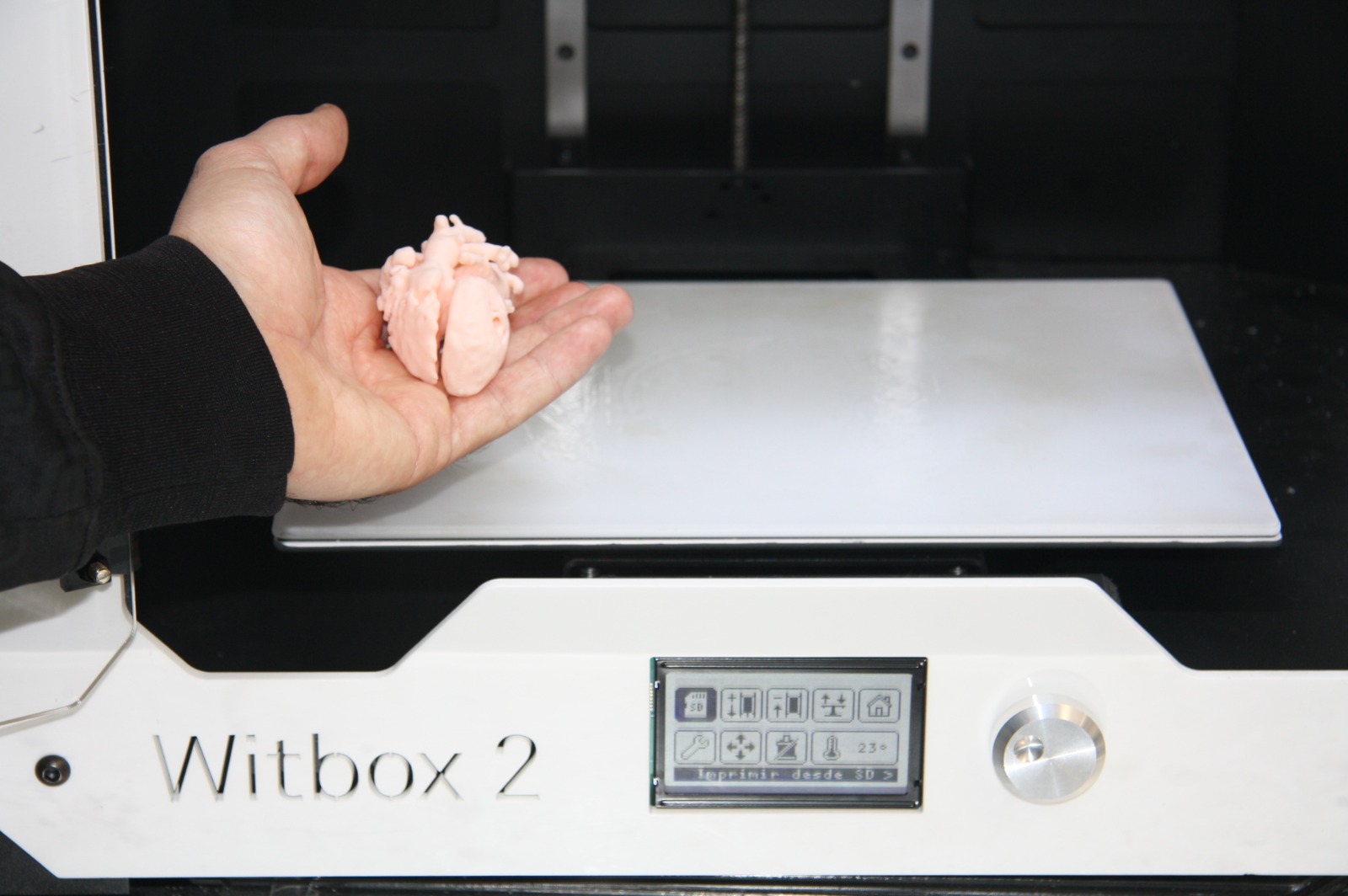
El servicio de diseño asistido por computador e impresión 3D, está destinado al soporte para la creación de nuevos prototipos, diseños o modelos anatómicos para los investigadores del centro.
El equipo dispone de una amplia experiencia en el diseño y fabricación de biomodelos y guías quirúrgicas de grado médico para la planificación de intervenciones, así como en el diseño y testeo de nuevos prototipos y diseños solicitados por los investigadores.
El laboratorio de fabricación (FABLAB HUVR-IBiS) dispone de diversas máquinas de impresión 3D tanto de filamento fundido como de resinas fotopolimerizables, incluyendo materiales de grado médico que pueden ser utilizados en quirófano.
Ubicación: 2ª Planta Edificio de Laboratorios Hospital Virgen del Rocío
Horario: Lunes a Viernes de 8:00 a 17:00 *
(*) Aquellos experimentos que requieran tiempo fuera del horario establecido se podrán realizar previo conocimiento del técnico responsable.

Dona
Fomenta la ciencia con una donación y forma parte del cambio.